Rib Cage Muscles Labeled - Thoracic Wall And Breast Illustrations
Rib Cage Muscles Labeled - Thoracic Wall And Breast Illustrations. In anatomy, the axis (from latin axis, axle) or epistropheus, is the second cervical vertebra (c2) of the spine, immediately posterior to the atlas, upon which the head rests. A rib has a flat body, as you can see from the picture of the anatomy of the human rib cage. The upper edge is round and the lower sharp. The thorax | basicmedical key. Sep 06, 2019 · there are 11 pairs of external intercostal muscles.
The upper edge is round and the lower sharp. The external intercostal muscles or rib cage muscles (located between the ribs) contract together with the diaphragm, lifting and expanding. The first seven ribs in the rib cage are attached to the sternum by pliable cartilages called costal cartilages; In most tetrapods, ribs surround the chest, enabling the lungs to expand and thus facilitate breathing by expanding the chest cavity. Diagram of human body, liver rib cage, rib cage diagram labeled, rib cage diagram numbered, rib cage diaphragm, rib cage heart, rib cage organs anatomy, rib cage pain, stomach, diagram of human body, liver rib cage, rib cage diagram labeled, rib cage diagram numbered, rib cage diaphragm, rib cage.
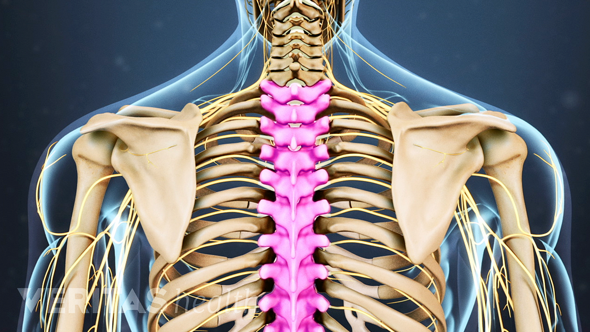
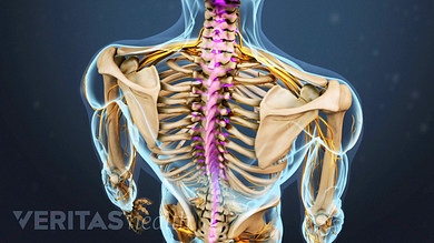
Thoracic Spine Anatomy And Upper Back Pain from embed.widencdn.net The thoracic cage is a component of the thoracic wall and encloses the majority of the structures of the respiratory system. The rectus abdominis runs between the ribs and the pubic bone and supports movements between the rib cage and the pelvis. The head only articulates with the body of the t1 vertebra and therefore only one articulatory surface is present. The rib cage consists of 24 ribs, 12 on either side, and it shields the organs of the chest, including the heart and the lungs, from damage. The ribs protect vital organs within the thoracic cage, and they also assist with breathing. Or, maybe you were a little too enthusiastic about that new exercise program, and the muscles between your ribs won. See more ideas about anatomy, rib cage anatomy, anatomy study. Anatomy of rib cage area / the thoracic cage anatomy and physiology i :
Rib cage anatomy numbered :
Rib cage pain can be associated with bruising, difficulty taking a deep breath, joint pain, and more. The first rib is the widest, shortest and has the sharpest curve of all the ribs. The heart and lungs are while the rib cage provides secondary protection to organs in a human's abdominal are. It forms the bony framework for breathing. Human ribs diagramnumbered / an inhalation is accomplished when the muscular diaphragm, at the floor of the thoracic cavity, contracts and flattens, while the contraction of intercostal muscles lift the rib cage up and out. Of the remaining five ribs, which are called false, the first three have their costal cartilages connected to the cartilage above them. Read more below to learn what may be causing your rib pain and when to seek treatment. Rib bone anatomy quiz for students taking anatomy and physiology! The head only articulates with the body of the t1 vertebra and therefore only one articulatory surface is present. In anatomy, the axis (from latin axis, axle) or epistropheus, is the second cervical vertebra (c2) of the spine, immediately posterior to the atlas, upon which the head rests. The rectus abdominis is an important postural muscle. There are 11 pairs of external intercostal muscles. Of all 24 ribs, the first seven pairs are often labeled as 'true.' these bones are.
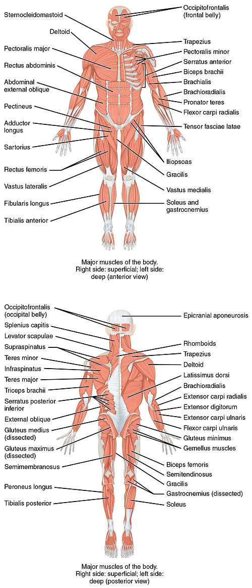
The ribs are attached to the breastbone, which is the. Diaphragm anatomy parts of the main structure the peripheral muscle. The major abdominal muscles include the transverse abdominals, the rectus abdominis, and the external and internal oblique muscles. Muscles of thorax, upper extremities, back and diaphragm are given connection by this cage. In spite of its resistance, the cage is dynamic, allowing pulmonary ventilation to.
Spinal Anatomy And Back Pain from embed.widencdn.net Of all 24 ribs, the first seven pairs are often labeled as 'true.' these bones are. It is responsible for pulling the rib cage toward the pelvis. They run inferoanteriorly from the rib above to the rib below, and are continuous with the external oblique of the abdomen. In anatomy, the axis (from latin axis, axle) or epistropheus, is the second cervical vertebra (c2) of the spine, immediately posterior to the atlas, upon which the head rests. The head only articulates with the body of the t1 vertebra and therefore only one articulatory surface is present. If the rib cage pain is due to a minor injury, such as a pulled muscle or bruise, you can use a cold compress on the. Apr 29, 2019 · the most common causes of rib cage pain are a pulled muscle or bruised ribs. In this rib bones anatomy quiz, you can test your knowledge of the ribs.
Possible causes of liver pain, or pain in the right side under the rib cage, include chronic hepatitis, liver abscess, fatty liver disease and liver.
These ribs are called true ribs. They run inferoanteriorly from the rib above to the rib below, and are continuous with the external oblique of the abdomen. Anatomy of rib cage area / the thoracic cage anatomy and physiology i : Or, maybe you were a little too enthusiastic about that new exercise program, and the muscles between your ribs won. Diagram of human body, liver rib cage, rib cage diagram labeled, rib cage diagram numbered, rib cage diaphragm, rib cage heart, rib cage organs anatomy, rib cage pain, stomach, diagram of human body, liver rib cage, rib cage diagram labeled, rib cage diagram numbered, rib cage diaphragm, rib cage. Diaphragm anatomy parts of the main structure the peripheral muscle. The major abdominal muscles include the transverse abdominals, the rectus abdominis, and the external and internal oblique muscles. Read more below to learn what may be causing your rib pain and when to seek treatment. It has a significant role play for raising or reducing the. The thoracic cage is a component of the thoracic wall and encloses the majority of the structures of the respiratory system. The rectus abdominis runs between the ribs and the pubic bone and supports movements between the rib cage and the pelvis. Muscles of thorax, upper extremities, back and diaphragm are given connection by this cage. It's nature is osteocartilaginous and elastic.
Human ribs diagramnumbered / an inhalation is accomplished when the muscular diaphragm, at the floor of the thoracic cavity, contracts and flattens, while the contraction of intercostal muscles lift the rib cage up and out. Or, maybe you were a little too enthusiastic about that new exercise program, and the muscles between your ribs won. It forms the bony framework for breathing. Read more below to learn what may be causing your rib pain and when to seek treatment. 16 photos of the rib cage diagram with organs.
List Of Skeletal Muscles Of The Human Body Wikipedia from upload.wikimedia.org The major abdominal muscles include the transverse abdominals, the rectus abdominis, and the external and internal oblique muscles. The ribs protect vital organs within the thoracic cage, and they also assist with breathing. Anatomy of rib cage area / the thoracic cage anatomy and physiology i : Muscle spasms felt within the rib cage may also be caused by the abdominal muscles. The rib cage consists of 24 ribs, 12 on either side, and it shields the organs of the chest, including the heart and the lungs, from damage. It forms the bony framework for breathing. The dome shaped thoracic cage provides the necessary rigidity for organ protection, weight support for the upper limbs and anchorage for muscles. Apr 29, 2019 · the most common causes of rib cage pain are a pulled muscle or bruised ribs.
Animated full human body anatomy.
Human ribs diagramnumbered / an inhalation is accomplished when the muscular diaphragm, at the floor of the thoracic cavity, contracts and flattens, while the contraction of intercostal muscles lift the rib cage up and out. With the upper ribs, closer to the nodule (and in the case of lower ribs, a little further from the nodule) they are curved and have a rough surface that connects them with muscles, angulus costae. Rib cage anatomy numbered : The rib cage consists of 24 ribs, 12 on either side, and it shields the organs of the chest, including the heart and the lungs, from damage. The rectus abdominis is an important postural muscle. Mar 20, 2015 · the human rib cage is made up of 12 paired rib bones; Of the remaining five ribs, which are called false, the first three have their costal cartilages connected to the cartilage above them. Aalso known as the six pack, is a paired muscle running vertically on each side of the front wall of the abdomen. The external intercostal muscles or rib cage muscles (located between the ribs) contract together with the diaphragm, lifting and expanding. This video covers the anatomy of the external, internal and innermost intercostal muscles, their origin, insertion, innervation and functions. The heart and lungs are while the rib cage provides secondary protection to organs in a human's abdominal are. Of all 24 ribs, the first seven pairs are often labeled as 'true.' these bones are. Or, maybe you were a little too enthusiastic about that new exercise program, and the muscles between your ribs won.
There are three parts of the peripheral muscle, sternal, costal, and lumbar, depending on the location of the peripheral attachment rib cage muscles. See more ideas about anatomy, rib cage anatomy, anatomy study.


No comments:
Post a Comment